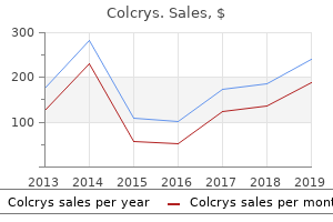
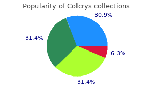
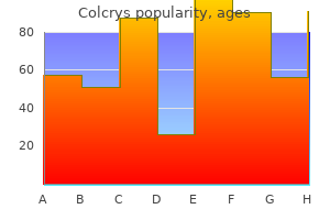
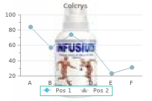
Colcrys
Order colcrys 0.5mg on-lineOnce parts are removed all cement debris does oral antibiotics for acne work generic 0.5mg colcrys, granulation tissue and delicate tissue is removed antibiotic cream for dogs generic colcrys 0.5 mg online. Once all elements are eliminated a sequence of events needs to be established taking into account the Select Appropriate Revision Implants It is important to assemble all of the implants earlier than cementing is initiated. Though it presents a good chance of eradicating the an infection, dependable pain reduction and a secure limb, the absence of knee motion makes it much less attractive to sufferers. Various fixation strategies are in use (external fixation, intramedullary nail, plates and screws) with 61% fusion price utilizing the external fixator and 95% fusion rate using a modular titanium intramedullary nail. Nonunion, malunion, limb shortening, nail migration, pin tract an infection, recurrent deep an infection and ipsilateral limb fractures are a few of the problems observed. Similarly if the gaps are loose both in extension and flexion rising the height of the insert could help. Here an offset stem extension directed posteriorly strikes the component anteriorly opening the flexion space. If the knee is tight in extension but stable in flexion further distal femoral cuts may be required. An offset stem extension directed anteriorly strikes the part posteriorly decreasing the flexion house. An undersized femoral element might have to be upsized to improve the posterior offset lowering the flexion gap. Present day modular hinged implants devices have a more conforming articulation permitting for the femoral condyles to interact with the tibial surface as the principle articulating surface have higher survivorship. Early weight bearing and knee mobilization is permitted after surgical procedure typically as early as the second or third postoperative days within the limits of pain. However in instances of V�Y quadricepsplasty or tibial tubercle osteotomy or extensive bone grafting immobilization as a lot as 3 weeks may be required for healing to occur. Complications include avulsion of the patellar tendon, fractures of the patella or tibia and femur throughout implantation, collateral Our Experience We retrospectively reviewed the outcomes of 142 sufferers, revised for septic and aseptic failures from January 2000 to December 2008. Our study revealed no statistical differences in the knee society scores and the Kaplan-Meier survivorships in each the teams. The use of a modified V�Y quadricepsplasty throughout total knee replacement to gain exposure and achieve flexion in the ankylosed knee. Osteotomy of the tibial tubercle during complete knee replacement: a report of 26 instances. Mid-to long-term results after therapy of 118 cases of periprosthetic infections after knee joint replacement utilizing one stage change surgery. Impaction grafting and wire mesh for uncontained defects in revision knee arthroplasty. San Francisco: proceedings of the 68 annual assembly of the American Academy of Orthopedic surgeons. Multiple irrigation, debridement and retention of parts in contaminated total knee arthroplasty. Infected total knee arthroplasty treated by arthroscopic irrigation and debridement.
Purple Sprouting Broccoli (Broccoli). Colcrys. - Are there safety concerns?
- Preventing prostate, breast, colon, rectal, bladder, and stomach cancer.
- Dosing considerations for Broccoli.
- How does Broccoli work?
- What is Broccoli?
Source: http://www.rxlist.com/script/main/art.asp?articlekey=97095
Buy colcrys 0.5mg mastercardIf the optic canal fails to develop norm ally infection vaginale purchase colcrys 0.5 mg with visa, it m ay compress and dam age the optic nerve infection questions cheapest generic colcrys uk, leading to visual eld defect s. All of the constructions depicted right here shall be thought of in m ore detail in subsequent pages. Anterior cranial fossa Middle cranial fossa Posterior cranial fossa Frontonasal pillar Anterior transverse pillar Pterygoid pillar Midlongitudinal pillar Vertical zygom atic pillar Horizontal zygom atic pillar b Foram en m agnum Posterior transverse pillar B the cranial fossae a Interior view, b midsagit tal part. The cranial fossae are bounded by the following constructions: � Anterior to m iddle: the lesser wings of the sphenoid bone and the jugum sphenoidale. In response to m asticatory pressures and different m echanical stresses, the bones of the cranium base are thickened to form "pillars" along the principal traces of drive (compare with the drive distribution in the anterior view on p. An analogous phenom enon of t ypical fracture traces is found within the m idfacial area (see the anterior views of LeFort fractures on p. Bones, Liga ments, a nd Joints Cribriform plate Frontal crest Frontal sinus Optic canal Anterior clinoid process Foram en rotundum (partially obscured) Foram en ovale Foram en spinosum Arterial groove Foram en lacerum Clivus Ethm oid bone, crista galli Frontal bone Sphenoid bone, lesser wing Sphenoid bone, higher wing Sphenoid bone, hypophyseal fossa Posterior clinoid course of Tem poral bone, petrous half Internal acoustic m eatus Jugular foram en Hypoglossal canal Groove for sigm oid sinus Foram en m agnum Cerebellar fossa Groove for transverse sinus Cerebral fossa D Interior of the bottom of the skull It is attention-grabbing to com pare the openings within the interior of the bottom of the skull with the openings seen in the external view (see p. An exam ple of this is the interior acoustic m eatus, through which the facial nerve, am ong different constructions, passes from the inside of the cranium into the petrous a part of the tem poral bone. Most of it s bers then go away the petrous bone by way of the st ylom astoid foram en, which is visible from the external aspect (see pp. The cribriform plate of the ethm oid bone connects the nasal cavit y with the anterior cranial fossa and is perforated by num erous foram ina for the passage of the olfactory bers (see p. Note: Because the bone is so thin in this area, a frontal head injury m ay simply fracture the cribriform plate and lacerate the dura m ater, allowing cerebrospinal uid to enter the nose. When pain is present within the distribution of the trigem inal nerve, tenderness to pressure m ay be famous at the supraorbital web site. The lateral orbital wall has been rem oved in b, and the m edial orbital wall has been rem oved in c. The orbit is kind ed by seven di erent bones (indicated right here by color shading): the frontal bone, zygom atic bone, m axilla, ethm oid bone, sphenoid bone (see a and c), and also the lacrim al bone and palatine bone, that are seen solely in the m edial view (see b). Frontal incisure Zygom aticoorbital foram en Nasolacrim al canal � Nasolacrim al duct 24 Head a nd Neck 2. Bones, Liga ments, a nd Joints Frontal incisure Supraorbital foramen Frontal bone, orbital part Zygom aticoorbital foram en Superior orbital fissure Zygom atic bone Inferior orbital fissure Infraorbital groove Posterior ethm oidal foram en Anterior ethm oidal foram en Optic canal Nasal bone Maxilla, frontal process Lacrim al bone Ethm oid bone, orbital plate a Maxilla, orbital surface Infraorbital foram en Frontal bone, orbital surface Lacrim al bone Anterior ethm oidal foram en Posterior ethm oidal foram en Ethm oid bone Optic canal Superior orbital fissure Foram en rotundum Inferior orbital fissure Maxilla, frontal course of Posterior lacrim al crest (lacrim al bone) Anterior lacrim al crest (m axilla) Fossa of lacrim al sac (with opening for nasolacrim al duct) Maxilla, orbital surface Infraorbital canal Maxillary hiatus Maxillary sinus Infraorbital foram en Frontal bone, orbital floor Frontal sinus Zygom atic bone orbital surface Zygom aticoorbital foram en Maxilla, orbital floor Infraorbital canal Inferior orbital fissure Superior orbital fissure b Pterygopalatine fossa Sphenoid bone, lesser wing Sphenoid bone, higher wing Maxillary sinus Palatine bone, pyram idal course of C Openings and pathw ays for neurovascular structures Right orbit, anterior view (a), lateral view (b), and m edial view (c). The following openings for the passage of neurovascular constructions (see itemizing in B) can be identi ed: the superior and inferior orbital ssures (a�c), the optic canal (a, b), the anterior and posterior ethm oidal foram ina (b, c), the infraorbital groove (a), which m erges into the infraorbital canal (b, c) and ends within the infraobital foram en (a, b); Supraobital foram en and frontal incisure (a); Zygom atico-orbital foram en (c). Diagram b reveals the ori ce of the nasolacrim al duct, by which lacrim al uid is conveyed to the inferior m eatus of the nose (see p. The inferior orbital ssure opens into the pterygopalatine fossa, which borders on the posterior wall of the m axillary sinus. It incorporates the pterygopalatine ganglion, an important element of the parasym pathetic nervous system (see pp.
Buy discount colcrys on-lineThe lat ter arch is the final to develop and is usually thought-about part of the fourth pharyngeal arch bacteria animation order generic colcrys from india. The exterior auditory canal is derived from the rst pharyngeal cleft bacteria kingdom characteristics cheapest colcrys, the t ym panic cavit y and eustachian tube from the rst pharyngeal pouch, and the palatine tonsil from the second pharyngeal pouch. Median cysts and stulas in the neck (a, b) are rem nant s of the thyroglossal duct. Lateral cysts and stulas within the neck are anom alous rem nant s of the ductal portions of the cervical sinus, which kind s because of tissue m igration throughout em bryonic developm ent. If epithelium -lined rem nant s persist, neck cyst s (right) or stulas (left) m ay appear in postnatal life (c). A full stula opens into the pharynx and onto the surface of the skin, whereas an incomplete (blind) stula is open at one end solely. The external ori ce of a lateral cervical stula is t ypically positioned on the anterior border of the sternocleidom astoid m uscle. This view was selected as an introduction to the cranium as a outcome of it displays the best num ber of cranial bones (indicated by di erent colours in B). The particular person bones and their salient features in addition to the cranial sutures and apertures are described in the models that follow. The chapter as a whole is intended to fam iliarize the reader with the nam es of the cranial bones before continuing to ner anatom ical particulars and the relationships of the bones to one another. Bones, Liga ments, a nd Joints Temporal bone, squam ous part Frontal bone Parietal bone Sphenoid bone, higher wing Ethm oid bone Lacrim al bone Nasal bone Zygom atic bone Maxilla Occipital bone Mandible Temporal bone, petrous part Temporal bone, t ympanic half B Lateral view of the cranial bones Left lateral view. The bones are shown in di erent colours to dem onstrate m ore clearly their extent s and limits. C Bones of the neurocranium (g ray) and viscerocranium (orange) Left lateral view. E Bones of the neurocranium and viscerocranium Neurocranium (gray) Viscerocranium (orange) � Front al bone � Sphenoid bone (excluding the pterygoid process) � Temporal bone (squam ous part, petrous part) � Pariet al bone � Occipit al bone � Ethm oid bone (cribriform plate) � Auditory ossicles � Nasal bone � Lacrim al bone � Ethmoid bone (excluding the cribriform plate) � Sphenoid bone (pterygoid process) � Maxilla � Zygom atic bone � Tem poral bone (t ym panic half, st yloid process) � Mandible � Vom er � Inferior nasal turbinate � Palat ine bone � Hyoid bone (see p. The bones of the cranium both develop directly from m esenchym al connective tissue (intram em branous ossi cation, gray) or form indirectly by the ossi cation of a cartilaginous m odel (enchondral ossi cation, blue). Elem ents derived from intram em branous and endochondral ossi cation (desm ocranium and chondrocranium respectively) m ay fuse collectively to form a single bone. This explains why congenital defect s of intram em branous ossi cation a ect each the skull and clavicle (cleidocranial dysostosis). F Bones of the desmocranium and chondrocranium Desmocranium (gray) Chondrocranium (blue) � � � � � � � � Nasal bone Lacrimal bone Maxilla Mandible Zygom atic bone Frontal bone Parietal bone Occipital bone (upper part of the squam a) � Temporal bone (squam ous half, t ympanic part) � Palatine bone � Vomer � Ethmoid bone � Sphenoid bone (excluding the medial plate of the pterygoid process) � Tem poral bone (petrous and m astoid half s, st yloid process) � Occipital bone (excluding the upper a part of the squam a) � Inferior nasal turbinate � Hyoid bone (see p. The bony m argins of the anterior nasal aperture m ark the start of the respiratory tract in the skull. The nasal cavit y, just like the orbit s, accommodates a sensory organ (the olfactory m ucosa). The anterior view of the cranium also shows the three clinically important openings via which sensory nerves cross to provide the face: the supraorbital foram en, infraorbital foram en, and m ental foram en (see p. Bones, Liga ments, a nd Joints Frontal bone Parietal bone Sphenoid bone, greater wing Nasal bone Ethm oid bone, m iddle nasal concha Inferior nasal concha Temporal bone Sphenoid bone, higher wing Zygom atic bone Maxilla Frontal sinus Ethm oid cells Sphenoid sinus Maxillary sinus Nasal cavit y Mandible B Cranial bones, anterior view Frontonasal pillar Horizontal zygom atic pillar C Paranasal sinuses: pneumatization lightens the bone Anterior view.
Order colcrys online nowElectrodiagnostic tests may show fibrillations and altered nerve conduction even at a number of days antibiotics for dogs uti buy colcrys overnight delivery. It should be confused that the pseudomeningoceles seen may not necessarily imply root avulsions in obstetrical palsies bacteria kit buy 0.5mg colcrys overnight delivery. At three weeks, the mom is instructed to mobilize the shoulder, elbow and hand. This is, notably, necessary in C56 palsies as the baby lacks strength within the abductors and external rotators whereas adduction and inner rotation is preserved. Similarly, the triceps needs to be stretched by full passive flexion of the elbow. In addition, the mother and father have to be instructed to stroke the palm and fingers repeatedly to provide sensory enter to the brain and to preserve awareness of the paralyzed higher limb. The clinician ought to be alert to detect incipient contractures in inside rotation. Instances of posterior dislocations of the shoulder have been famous even in the early levels. This represents 46% of all instances and is related to the most favorable prognosis. It is a delicate matter to explain the mechanism of damage without, indirectly, blaming the obstetrician. The clinician must resist the temptation to prescribe a splint or any investigations at such an early stage. In most circumstances of C56 palsies, abduction returns quickly and the child can take the arm to 90� in the plane of the trunk within a month. It is simple to be fooled into believing that the child has recovered biceps perform. This can be inferred only when the child flexes the elbow from the absolutely prolonged place with the arm by the facet of the trunk. Cases with more in depth deficits at start may present recovery of the triceps and pectoralis main in the first two months. Prognosis and Role of Nerve Reconstruction Surgery There is no doubt that an in depth deficit implies a extreme lesion and that the nerves are torn. In such patients, one should offer surgical procedure at the acceptable time to direct the rising ends of the torn nerves in the direction of the particular targets that may allow restoration of maximal functions. Evaluation of recovery in such children is often challenging, as mentioned earlier. The "towel take a look at," described by Bertelli, is a helpful tool to stimulate the child to try to attain the face and, thus, reveal presence or absence of restoration. The focus is on restoration of the biceps but this has to be seemed for within the context of overall return of shoulder and elbow functions. Clarke and his associates14 have described their technique of evaluation of restoration in each of the misplaced functions. They advocate early surgery in sufferers with evident root avulsions whereas different kids are reviewed periodically.
Buy colcrys paypalWhen the m ucosa (ciliated respiratory epithelium) in the ethm oid cells (green) becom es swollen because of antimicrobial natural purchase genuine colcrys line in am m ation (sinusitis) infection vaginal order colcrys australia, it blocks the ow of secretions (see arrows) from the frontal sinus (yellow) and m axillary sinus (orange) in the ostiom eatal unit (red). Because of this blockage, m icro- organism s also becom e trapped in the other sinuses, the place they m ay incite in am m ation. Thus, while the anatom ical focus of the illness lies within the ethm oid cells, in am m atory symptom s are also m anifested in the frontal and m axillary sinuses. In affected person s with persistent sinusitis, the slender websites can be surgically widened to establish an e ective drainage route, alleviating the condition. It type s the bony housing for the auditory and vestibular equipment and bears the articular fossa of the temporom andibular joint. The temporal bone develops from three facilities that fuse to form a single bone: � the squam ous part, or temporal squam a (light green), bears the articular fossa of the temporom andibular joint (m andibular fossa). Note: the st yloid course of seems to belong to the t ympanic a half of the tem poral bone because of it s location. Chorda t ym pani Facial nerve Mastoid air cells Tympanic m em brane Pharyngot ym panic (auditory) tube Internal carotid artery Internal jugular vein Mastoid process C Projection of clinically necessary buildings onto the left temporal bone the t ympanic m em brane is shown translucent in this lateral view. Because the petrous bone incorporates the m iddle and internal ear and the t ym panic m em brane, a information of it s anatomy is of key significance in otological surgical procedure. The internal floor of the petrous bone has openings (see D) for the passage of the facial nerve, inner carotid artery, and inner jugular vein. A sm all nerve, the chorda t ympani, passes by way of the t ympanic cavit y, and lies m edial to the t ympanic m em brane. The chorda t ympani arises from the facial nerve, which is vulnerable to harm throughout surgical procedures (cf. The m astoid strategy of the petrous bone kind s air- lled cham bers, the m astoid cells, that fluctuate greatly in size. Because these cham bers com m unicate with the m iddle ear, which in flip com m unicates with the nasopharynx via the pharyngot ympanic (auditory) tube (also referred to as eustachian tube) micro organism within the nasopharynx m ay move up the pharyngot ym panic tube and achieve access to the m iddle ear. From there they m ay cross to the m astoid air cells and nally enter the cranial cavit y, inflicting m eningitis. Bones, Liga ments, a nd Joints Zygom atic course of Temporal surface External acoustic opening Articular tubercle Mandibular fossa Petrot ym panic fissure St yloid process Mastoid course of Mastoid foram en External acoustic m eatus Tympanom astoid fissure Tympanosquam ous fissure St yloid process Zygom atic course of a Articular tubercle Mandibular fossa Arterial groove Carotid canal Petrot ympanic fissure Jungular fossa St ylom astoid foram en External acoustic opening Mastoid process Mastoid notch Petrous pyram id b Zygom atic process Mastoid foram en Mastoid foram en Petrous apex Internal acoustic m eatus c Groove for sigm oid sinus St yloid process D Left temporal bone a Lateral view. The m astoid process develops progressively in life due to traction from the sternocleidom astoid m uscle and is pneum atized from the inside (see C). The shallow articular fossa of the temporom andibular joint (the mandibular fossa) is clearly seen from the inferior view. The facial nerve em erges from the base of the cranium via the st ylom astoid foram en.
Order colcrys with american expressNonunions can Conservative Management of Fractures Adequate immobilization within the type of lightweight splints or casts is appropriate antibiotics for acne scars buy colcrys 0.5mg online. If the fracture is secure after a few weeks antibiotic resistance yersinia pestis order generic colcrys, detachable splints or orthoses that provide enough stability may be used, so that the limb can be protected throughout weight-bearing activities. The lower extremities are typically concerned to a higher extent than the upper extremities functionally. Surgical remedy at a younger age could require revision before surgery carried out in an older youngster. This relative disadvantage is clearly countered by the enhanced progress, growth and quality of life gained by way of early surgical stabilization. Preoperative Anesthesia Considerations Preoperative evaluation is crucial, including evaluation for cranial cervical abnormalities. Stabilization of the neck by the surgeon assisting anesthesiology during intubation is critical. The whole operative staff, as well as the preoperative and postoperative nursing employees, must be educated in the care, monitoring, and handling of these youngsters to forestall fractures. Postoperative pain administration can be challenging, as many of the children have been uncovered to ache medications throughout their lives. Spasm is often a major component of postoperative discomfort and short-term, low-dose diazepam can be quite beneficial. They predictably fail and predispose to recurrent fractures on the ends of the plates. The young kids are generally handled with common non-telescoping rods (K-wires, Rush pins, elastic rods) can be used. In the upper extremities, the situation is different: the humeri could be rodded with telescopic rods (much more difficult than decrease extremity 3141 rodding) or with common rods (either from the top-down, or from the bottom-up-"Eiffel Tower" construct). One of the primary advantages of this technique is that a quantity of bones may be handled on the same surgical setting, limiting the variety of surgical procedures and reducing the rehabilitation that these kids endure. So far, due to the bone fragility, titanium rods (elastic rods or Nancy nails) appear to be too elastic and the tutor effect of the rod is kind of ineffective as a outcome of the pliability of the nail. When used as a "sliding rod" system, it loses the profit of the "belly" and the mechanical stability is compromised. The sagittal airplane is the most important diameter to contemplate when choosing the dimensions of the implant. They have traditionally been used in conjunction with intensive surgical approaches, and require that an arthrotomy of the knee be performed for insertion within the femur and tibia, and an extensive arthrotomy of the ankle to insert this gadget within the tibia. With progress, bowing in the central segment of the rodded bone can happen with transcortical migration of the rods and in the end fracture.
Syndromes - Bleeding
- Bones of the spine
- Wearing away of materials placed during surgery, such as a sling or artificial sphincter
- ECG
- Biopsy of the muscle, organ, tissue, or nerve biopsy
- Elavil
- Eye pain
- Irritability
- To get to the lungs, blood must cross an atrial septal defect (ASD), ventricular septal defect (VSD), or a patent ductus artery (PDA). (These conditions are described above.) This condition severely restricts blood flow to the lungs.
Order 0.5 mg colcrys fast deliveryIf this flap will get shredded or is poorly developed antibiotic for dog uti buy colcrys 0.5 mg otc, good watertight closure is difficult antibiotics for acne results order colcrys 0.5mg mastercard. If that is inadequate, use a curved sharp osteotome to resect the posteromedial osteophyte without damaging the medial collateral ligament. The entry hole is made and over-reamed to accept the medullary canal rod of the jig. The angle of resection is judged on the lengthy leg scanogram accomplished in neutral rotation, and is normally set 5�7� to get cut perpendicular to mechanical axis. The slicing block is fixed to the anterior side of femur with minimum three pins and the jig is taken off. The distal femur is cut with oscillating saw to resect 9�10 mm of distal femur on much less worn side. The noticed must be held firmly with the flat floor of the noticed blade stored flush with the distal face of cutting slot. This spike rests of the distal femur and it is very important cushion it with a surgical cotton mop. The jig is first positioned across the ankle and rotation of the jig is aligned to the junction of medial third and lateral two third of the tibial tubercle. Proximally, the jig could have a spike that may be fastened to the center of the tibia. Most methods have the slope marked on the slicing block and the anterior arm of the jig is stored parallel to the shin of tibia. The heart of the ankle joint is medial to the mid-malleolar level and the place of the jig is about accordingly. Other reference landmarks that can be used include tibialis anterior tendon and the second metatarsal. However, in presence of foot deformities or a very mobile foot joints, these landmarks are unreliable. After the tibial jig is locked in position, the slope of the reduce is checked using angel wing depth resection gauge. In a nondeformed varus knee, 9�10 mm is resected using the lateral facet as reference. It is important not to use extra-long chopping noticed blade as it could possibly go beyond the tibia to injure vital structures. The knee is then positioned in extension and remaining medial meniscus is excised taking care not to damage the medial collateral ligament. Extension Gap With knee in extension, extension gap is first visually checked and balance is then checked with spacer blocks.
Order cheap colcrys lineBruises on back of head bacteria staphylococcus aureus discount colcrys 0.5mg without a prescription, neck antibiotics dogs purchase 0.5mg colcrys with amex, legs and arms, on the buttocks, abdomen, cheeks or genitalia could also be suspicious for abuse. When an toddler with musculoskeletal damage presents with altered mental status, whiplash shaken infant syndrome ought to be suspected. Violent shaking of a small child whiplashes is relatively massive head back and forth over the thorax with potential improvement of subdural hematomas. Multiple fractures,7 evidence of repeated trauma, bruises and lacerations confirm the prognosis. The repetitive nature of the damage is hallmarked by the presence of various levels of bone restore. The Skeletal Survey A skeletal survey is helpful for finding out additional fractures in battered kids. Radiologic Features There is always risk of multiple skeletal lesions, which is nicely described by Silverman in 1953. He offered a report on a number of long bone fractures without subdural hematoma in three kids and established their traumatic basis. There is all the time predilection for the metaphysis with exaggerated periosteal response and multiplicity of the lesions in numerous levels of therapeutic and restore. The physeal injuries with gross or minimal displacement of the epiphysis are frequent. There is profuse subperiosteal new bone formation, which converts into thick cortex Diagnosis the child with a number of fractures, proof of repeated trauma, bruises and lacerations normally raises suspicion of battered child syndrome. The radiogram reveals the presence of various stages of bone repair, which is the hallmark of kid abuse. Differential Diagnosis In the differential prognosis, one should rule out osteogenesis imperfecta, congenital insensitivity to pain, infantile cortical hyperostosis, scurvy, congenital syphilis, Caffey illness, osteomy elitis, septic arthritis, fatigue fracture, osteoid osteoma and different tumors, rickets, leukemia, bleeding problems, hypophosphatasia, neuromuscular disease, metastatic neuroblastoma, osteopetrosis, kinky hair syndrome, prostaglandin remedy. It is essential to take the accountable members of the family into confidence and inform about this syndrome. The person concerned with battering the kid should be submitted to psychiatric remedy. Multiple fractures within the long bones of infants affected by continual subdural hematoma. National Center on Child Abuse and Neglect: National baby abuse and neglect Data System: Working paper 21991. Another methodology is mother or father training in particular parenting skills corresponding to primary childcare, self-discipline, child development and education and familiarity with local support services. Arthrography in the diagnosis of fractures of the distal finish of the humerus in infants.
Discount colcrys onlineAn further part is the alveolar bone virus 16 purchase colcrys with mastercard, which kind s a part of the alveolar pocket (socket) infection vaginal buy 0.5 mg colcrys otc. The alveolar sockets resem ble cups with numerous holes in their bony walls, the cribriform layer of bone. Blood and lymphatic vessels enter the periodontal ligam ent via these holes into the desmodontal gap where they type a dense lat ticework surrounding the dental roots. Note: Deciduous enamel are given Rom an num erals and the perm anent tooth Arabic num bers. Knowledge of the eruption pat tern is clinically essential since corresponding information helps to diagnose growth delay in youngsters. The anterior bone lam ella above the roots of the deciduous enamel has been removed, the underlying perm anent teeth are visible. A six year old was chosen as a result of at that age all deciduous tooth have erupted and are all nonetheless present. Y at the sam e tim e, the anteet, rior m olar has started to erupt as the rst perm anent tooth (see C). Second perm anent prem olar First perm anent prem olar Perm anent canine a Interm axillary suture Second deciduous m olar First deciduous m olar Perm anent lateral incisor Perm anent central incisor Deciduous lateral incisor Deciduous canine Deciduous canine Deciduous Deciduous central incisor lateral incisor First deciduous m olar Second decidous m olar First perm anent m olar Second perm anent m olar Second perm anent prem olar Mental foram en First perm anent prem olar Perm anent central incisor Perm anent lateral incisor Perm anent canine b Perm anent canine Perm anent lateral incisor First perm anent prem olar Deciduous central incisor Second perm anent m olar Second perm anent prem olar First perm anent m olar Second deciduous m olar c Deciduous Deciduous First decilateral incisor canine duous m olar Deciduous canine Deciduous lateral incisor Perm anent central incisor Perm anent lateral incisor Second perm anent m olar Second perm anent prem olar Perm anent canine First perm anent prem olar Second deciduous m olar First deciduous m olar First perm anent m olar d 53 Hea d and Neck 2. Localized epithelial thickening presents the rst m orphologically veri able signal of the beginning of tooth developm ent. They run in a horseshoe-shape parallel to the lip line and develop into the m esenchym e of the m axilla and m andible of a ve-week old hum an em b ryo (cf. In m esial-distal path, the free m argins on either side of the overall dental lam ina thickens to type 5 tooth buds every, equal to the ten prim ary teeth in each decrease and higher jaw. Subsequently, each of these tooth epithelial buds rework s rst into cap-shaped and later bell-shaped enam el organs (cf. Early Cap stag e: Bud- and cap-shaped collections of cells develop on account of intensive cell proliferation in the odontogenic epithelium. Their concavit y deepens on the far facet of the epithelium and starting from the m argin they develop across the m esenchym e (see C). Late Cap stage: � the enam el organ is com posed of an inside and outer enam el epithelium and the stellate reticulum, which lies in guess ween. The cells of the internal enam el epithelium grow increasingly colum nar-shaped on the basal lamina significantly across the enam el knot. Increasing extracellular m atrix production (stellate reticulum) leads to additional separation of the outer and inside enam el epithelium layers. Note: the perm anent tooth (m olars of the perm anent dentition), which are positioned distally from the prim ary dentition outcome from the dental lam ina, which elongates distally. Bell stage: � the stellate reticulum becom es more and more m ore volum inous and divides into a free m id-zone (stratum reticulum proper) and a cellular layer (stratum interm edium) im m ediately next to the internal enam el epithelium. Blood vessels and nerve bers develop into the dental papilla where the dental pulp later develops.
Order generic colcrys canadaIf the final reamer used is of dimension 50 mm anti virus 0.5 mg colcrys, then the 48 mm trial cup ought to be used so as to have enough layer of cement mantle antibiotics for uti without sulfa 0.5mg colcrys free shipping. Thorough pulsatile lavage must be used to take away delicate tissue, fat and loose bone. At this juncture the anesthetist should present hypotensive anesthesia however central venous stress should be enough. All pressurization methods have to be used to pressurize the cement into cancellous bone which will act as a micro-interlock. The acetabular component is then driven with Acetabular and Femoral Preparation After dislocation of the hip, measure the horizontal offset which is the space from pyriformis fossa to the middle of the pinnacle, which ought to be recreated with the synthetic joint. The acetabular strain must be sustained after proper orientation for anteversion and inclination. The beneficial inclination is between 35� and 45� to the horizontal plane and anteversion 15�20�. This is possible with inclination information and by making use of transverse acetabular ligament. Excessive cement must be removed with a blunt instrument to avoid harm to the polyethylene. The extreme osteophytes across the cup must be eliminated to avoid impingement and dislocation. All gentle tissues are cleared from the pyriformis fossa and the gluteus medius is retracted. In cemented arthroplasty removal of calcar femoralis is essential, to put the femoral component into the middle of the canal in lateral anatomical profile. Few checks for stability done at this level are� � Combined anteversion take a look at of Richard Scott and Ranawat- maintain the leg in a straight line with torso and rotate the hip internally till the head becomes coplanar with the cup. If "C" stem, Exeter stem or any extremely polished stem is used the block is used just on the tip of the stem. Proximal pressurization ought to be accomplished on the doughy consistency of the cement and gradually the vent should be eliminated. The final prosthesis is inserted from the trochanteric fossa preserving the thumb on the medial aspect to avoid cement extrusion. The prosthesis ought to be inserted progressively with out hammering so that adequate strain is created into the canal. The final degree of seating is identified and all cement is removed from the calcar space. Ankylosing Spondylitis Ankylosing spondylitis is a member of a gaggle of rheumatic diseases that affect the mobility of the backbone, collectively known as spondyloarthropathies. The illness is characterised by axial skeletal involvement (sacroilitis, spines, ribs and sternum) and likewise involvement of peripheral joints (hip, shoulder, hands and feet). Peripheral main joint arthritis can happen quickly after medical presentation of axial skeletal disease.
|
